Presentation Health and Disease Biology Diagrams The superior chambers consist of the right atrium and left atrium (plural, atria: L., corridor). which lie primarily on the posterior side of the heart.[Interior view/ Posterior view]Have you been making any of the common anatomy learning mistakes? Find out!
.PNG)
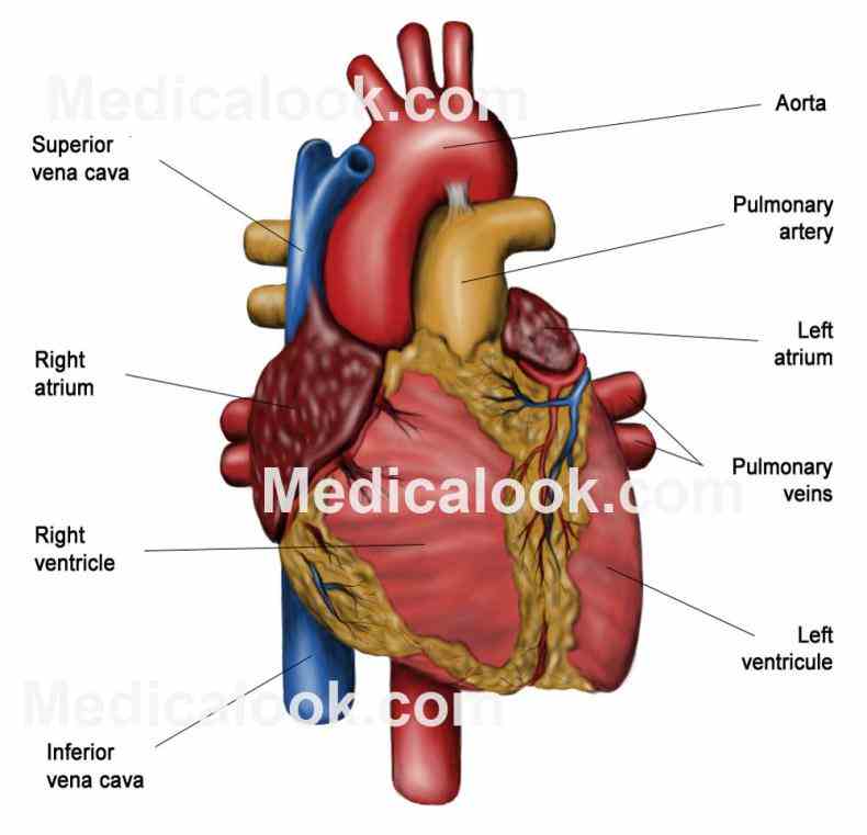
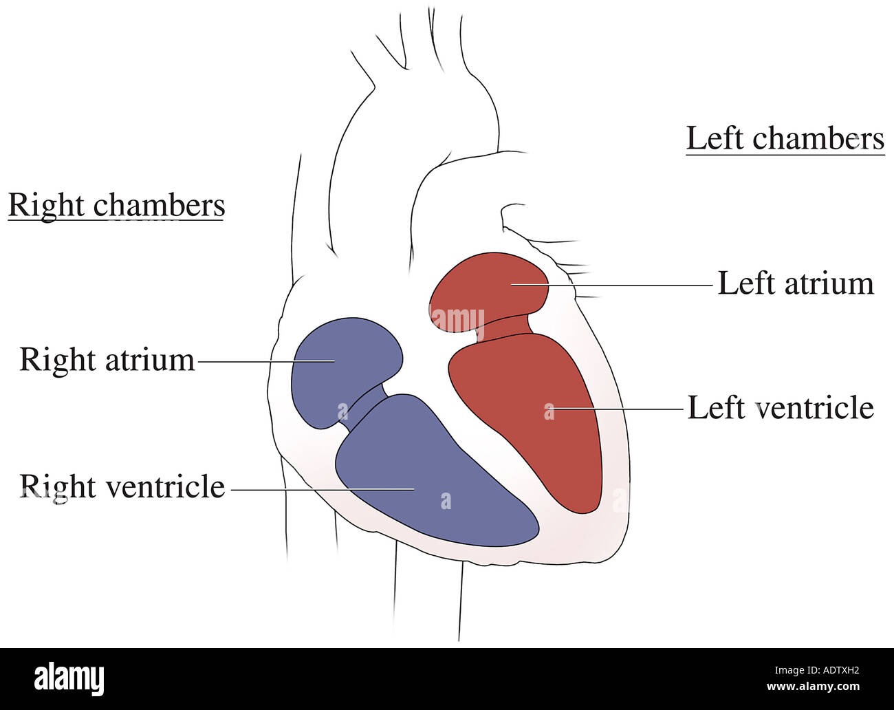
Anatomy of the interior of the heart. This image shows the four chambers of the heart and the direction that blood flows through the heart. Oxygen-poor blood, shown in blue-purple, flows into the heart and is pumped out to the lungs. Then oxygen-rich blood, shown in red, is pumped out to the rest of the body, with the help of the heart valves.
.jpg)
Chambers of the Heart Biology Diagrams
What are the heart chambers? Your heart chambers are four hollow spaces within your heart. There are two atria (upper chambers) called your right atrium and left atrium. In addition, there are two ventricles (lower chambers) called your right ventricle and left ventricle. Each chamber plays an important role in your heart's functioning.
+and+ventricles+(lower+chambers).jpg)
Understanding Heart Failure With Anatomy . Heart failure can result from various conditions that weaken or damage the heart muscle, impairing its ability to pump blood effectively. This can lead to a backup of blood in the heart's chambers or the blood vessels leading to the heart.
Chambers of the Heart Biology Diagrams
The Chambers of the Heart. The Conducting System of the Heart. The Heart Wall. The Pericardium. The Surfaces and Borders of the Heart. TeachMeAnatomy. Part of the TeachMe Series. The medical information on this site is provided as an information resource only, and is not to be used or relied on for any diagnostic or treatment purposes. The human heart consists of four chambers: The left side and the right side each have one atrium and one ventricle. Each of the upper chambers, the right atrium (plural = atria) and the left atrium, acts as a receiving chamber and contracts to push blood into the lower chambers, the right ventricle and the left ventricle.
Learn faster Surface anatomy and chambers of the heart Start quiz Heart valves Heart valves separate atria from ventricles, and ventricles from great vessels. The valves incorporate two or three leaflets (cusps) around the atrioventricular orifices and the roots of great vessels.