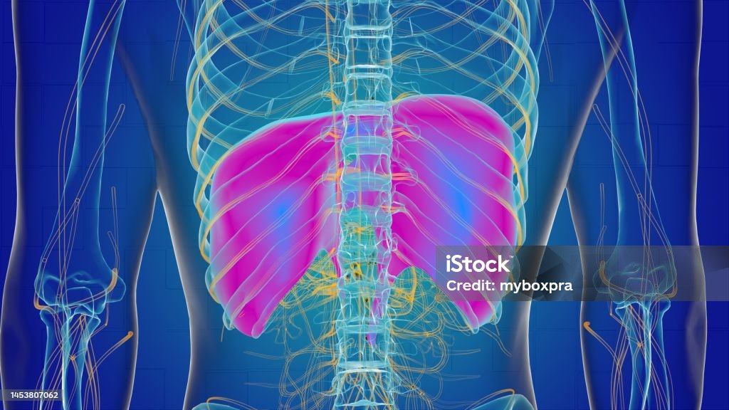
Human Diaphragm Anatomy for Medical Concept 3D Rendering Stock Biology Diagrams The diaphragm separates the thoracic cavity from the abdominal cavity. It is a double-domed, musculotendinous partition that consists of a continuous sheet of muscle surrounding a central tendon. The peripheral muscle is named according to its peripheral points of attachment. The sternal part attaches to the posterior aspect of the xiphoid process. The coastal part attaches to the internal

Anatomy . The diaphragm is a fibrous muscle that runs between the chest and abdomen, separating these two large cavities. It is asymmetric, as its right dome is larger than the left dome. The diaphragm has openings that allow certain structures to span the chest and abdominal cavities.
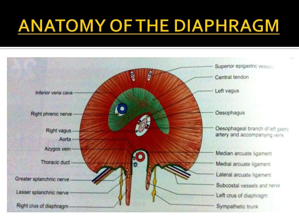
Diaphragm: Location, anatomy, innervation and function Biology Diagrams
Structure of diaphragm shown using a 3D medical animation still shot. The thoracic diaphragm, or simply the diaphragm (/ ˈ d aɪ ə f r æ m /; [1] Ancient Greek: διάφραγμα, romanized: diáphragma, lit. 'partition'), is a sheet of internal skeletal muscle [2] in humans and other mammals that extends across the bottom of the thoracic cavity.The diaphragm is the most important muscle
The diaphragm is a muscular partition that separates the thoracic cavity from the abdominal cavity in the human body. It is an important structure that plays a crucial role in respiration, as it is responsible for generating the negative pressure that helps to draw air into the lungs. Structure Anatomy The diaphragm is a dome-shaped
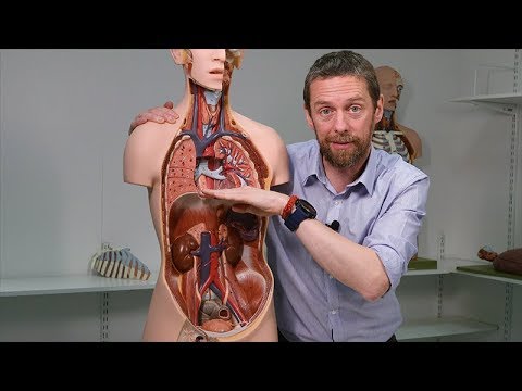
Diaphragm (Human Thorax) Location, Anatomy, Function and Position Biology Diagrams
The diaphragm in the thorax is called the thoracic diaphragm and serves as an important anatomical landmark that separates the thorax, or chest, from the abdomen. It functions during breathing when it contracts to enlarge the thoracic cavity and reduce the intrathoracic pressure so that lungs may expand and fill their alveoli with air. It is a dome-shaped muscle and tendon that functions as
